Montwood Medical Center has just installed a CT Scan (better known as a Cat Scan) for our patients, we know that it can be a big burden to pay for medical bills especially for services such as CT scans, so we lowered our base price to below half of what you can expect to pay at the hospitals. So if you need this special test we are ready to save you money!.
FAQ's :-
What is a CT Scan?
A computerized axial tomography scan is more commonly known by its abbreviated name, CAT scan or CT scan. It is an x-ray procedure which combines many x-ray images with the aid of a computer to generate cross-sectional views and, if needed, three-dimensional images of the internal organs and structures of the body. A CAT scan is used to define normal and abnormal structures in the body and/or assist in procedures by helping to accurately guide the placement of instruments or treatments. A large donut-shaped x-ray machine takes x-ray images at many different angles around the body. These images are processed by a computer to produce cross-sectional pictures of the body. In each of these pictures the body is seen as an x-ray "slice" of the body, which is recorded on a film. This recorded image is called a tomogram. "Computerized Axial Tomography" refers to the recorded tomogram "sections" at different levels of the body.
Imagine the body as a loaf of bread and you are looking at one end of the loaf. As you remove each slice of bread, you can see the entire surface of that slice from the crust to the center. The body is seen on CAT scan slices in a similar fashion from the skin to the central part of the body being examined. When these levels are further "added" together, a three-dimensional picture of an organ or abnormal body structure can be obtained.
Why are CT Scans performed?
CAT scans are performed to analyze the internal structures of various parts of the body. This includes the head, where traumatic injuries, (such as blood clots or skull fractures), tumors, and infections can be identified. In the spine, the bony structure of the vertebrae can be accurately defined, as can the anatomy of the intervertebral discs and spinal cord. In fact, CAT scan methods can be used to accurately measure the density of bone in evaluating osteoporosis.
Occasionally, contrast material (an x-ray dye) is placed into the spinal fluid to further enhance the scan and the various structural relationships of the spine, the spinal cord, and its nerves. CAT scans are also used in the chest to identify tumors, cysts, or infections that may be suspected on a chest x-ray. CAT scans of the abdomen are extremely helpful in defining body organ anatomy, including visualizing the liver, gallbladder, pancreas, spleen, aorta, kidneys, uterus, and ovaries. CAT scans in this area are used to verify the presence or absence of tumors, infection, abnormal anatomy, or changes of the body from trauma.
The technique is painless and can provide extremely accurate images of body structures in addition to guiding the radiologist in performing certain procedures, such as biopsies of suspected cancers, removal of internal body fluids for various tests, and the draining of abscesses which are deep in the body. Many of these procedures are minimally invasive and have markedly decreased the need to perform surgery to accomplish the same goal.
How do I prepare for a CT Scan?
In preparation for a CAT scan, patients are often asked to avoid food, especially when contrast material is to be used. Contrast material may be injected intravenously, or administered by mouth or by an enema in order to increase the distinction between various organs or areas of the body. Therefore, fluids and food may be restricted for several hours prior to the examination. If the patient has a history of allergy to contrast material (such as iodine), the requesting physician and radiology staff should be notified. All metallic materials and certain clothing around the body are removed because they can interfere with the clarity of the images.
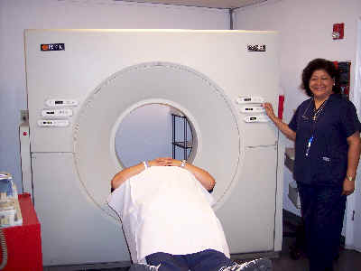
How is a CT Scan performed?
Patients are placed on a movable table, and the table is slipped into the center of a large donut-shaped machine which takes the x-ray images around the body. The actual procedure can take from a half an hour to an hour and a half. If specific tests, biopsies, or intervention are performed by the radiologist during CAT scanning, additional time and monitoring may be required. It is important during the CAT scan procedure that the patient minimize any body movement by remaining as still and quiet as is possible. This significantly increases the clarity of the x-ray images. The CAT scan technologist tells the patient when to breathe or hold his/her breath during scans of the chest and abdomen. If any problems are experienced during the CAT scan, the technologist should be informed immediately. The technologist directly watches the patient through an observation window during the procedure and there is an intercom system in the room for added patient safety.