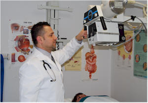
X-rays - Identify bony structures, X-rays have been developed for their use in medical imaging. Radiology is a specialized field of medicine. Radiographers employ radiography and other techniques for diagnostic imaging. Indeed, this is probably the most common use of X-ray technology. X-rays are especially useful in the detection of pathology of the skeletal system, but are also useful for detecting some disease processes in soft tissue. Some notable examples are the very common chest X-ray, which can be used to identify lung diseases such as pneumonia, lung cancer or pulmonary edema, and the abdominal X-ray, which can detect ileus (blockage of the intestine), free air (from visceral perforations) and free fluid (in ascites). In some cases, the use of X-rays is debatable, such as gallstones (which are rarely radiopaque) or kidney stones (which are often visible, but not always). Also, traditional plain X-rays pose very little use in the imaging of soft tissues such as the brain or muscle. Imaging alternatives for soft tissues are computed axial tomography (CAT or CT scanning), magnetic resonance imaging (MRI) or ultrasound.
 Ultrasounds - An ultrasound machine creates images that allow various organs in the body to be examined. The machine sends out high-frequency sound waves, which reflect off body structures. A computer receives these reflected waves and uses them to create a picture. Unlike with an x-ray, there is no ionizing radiation exposure with this test. Common ultrasounds are for pregnancy or obstetrics ultrasound, vascular ultrasound to check blood flow like in the heart or arms and legs called Doppler, abdominal ultrasounds and thyroid ultrasounds.
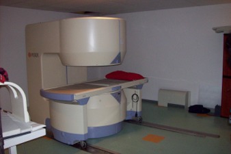 OPEN MRI - A magnetic resonance imaging, is a routine diagnostic procedure. Images of the internal tissues of the human body are produced by using the tiny magnets of hydrogen nuclei which are abundant in all of us as parts of water, fat, protein, and other molecules. The large magnetic field of the MRI machine causes the hydrogen magnets to align, while applied radio waves excite them to transmit signals similar to the radio waves generated at FM radio stations. The locations of the signals within the body are identified using magnetic field gradient pulses which are the source of the loud knocking noises heard during the examination. Once enough signals have been collected, they are processed by powerful computers to generate pictures of the human anatomy in vivid detail for the radiologist to analyze and diagnose abnormalities. For complete details see MRI. view more...
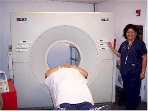 CT-Scan - A computerized axial tomography scan is more commonly known by its abbreviated name, CAT scan or CT scan. It is an x-ray procedure which combines many x-ray images with the aid of a computer to generate cross-sectional views and, if needed, three-dimensional images of the internal organs and structures of the body. A CAT scan is used to define normal and abnormal structures in the body and/or assist in procedures by helping to accurately guide the placement of instruments or treatments. A large donut-shaped x-ray machine takes x-ray images at many different angles around the body. These images are processed by a computer to produce cross-sectional pictures of the body. In each of these pictures the body is seen as an x-ray "slice" of the body, which is recorded on a film. This recorded image is called a tomogram. "Computerized Axial Tomography" refers to the recorded tomogram "sections" at different levels of the body. For complete details see CT Scan. view more...
 Bone Density - Bone density is a medical term referring to the amount of matter per cubic centimeter of bones. It is measured by a procedure called densitometry. No longer is it necessary to go to radiology or nuclear medicine departments of hospitals or clinics, because Montwood Medical Centers can perform done density scan in-house. The measurement is painless and non-invasive and involves minimal radiation exposure. Measurements are most commonly made over the lumbar spine and over the upper part of the hip. The forearm is scanned if either the hip or the lumbar spine can't be. For complete details see Bone Density.